×
模态框(Modal)标题
在这里添加一些文本
Close
Close
Submit
Cancel
Confirm
×
模态框(Modal)标题
×
Please choose a citation manager
RIS (ProCite, Reference Manager)
BibTeX
Content to export
Citation
Citation and abstract
Export
Author Login
Review Login
Office Work
Reader Login
Toggle navigation
Home
About JFBI
About JFBI
Editorial Board
For Readers
Accepted
Current Issue
All Issues
Most Read Papers
Most Downloaded Papers
For Authors
Scope of the JFBI
Author Login
Publication Fee
Paper Format Requirements
Call for Papers
For Reviewers
Instruction for Reviewer
Reviewer Regististration
Review Form
Reviewer Login
Call for reviewers
Related Information
For Editors
Instruction for Editor
Call for Editors
Editor Login
Download Forms
Service
Professional English Editing Service
Other Services
Related Information
Contact Us
Announcement
More >>
Current Issue
Volume 18 Issue 4
More
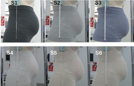
Comparison of Support and Clothing Pressure Distribution in Japanese and Australian Maternity Support Garments
Tamaki Takada-Mitsuno, Carolina Quintero Rodriguez
View
Design Guidelines for Accessible Dining Apparel for the Visually Impaired: Toward Inclusive and Sustainable Fashion
Yu-He Qin, Zi-Rui Ji, Jun-Xian Li, Jia-Yi Lin, Kai-Yue Yu, Jing Guo
View
Natural Mordants in Focus: A Comparative Study of Alum and Calcium Hydroxide in Cotton Dyeing with Senna Siamea Heartwood
Piyanut Jingjit, Yanisa Komonsirichok
View
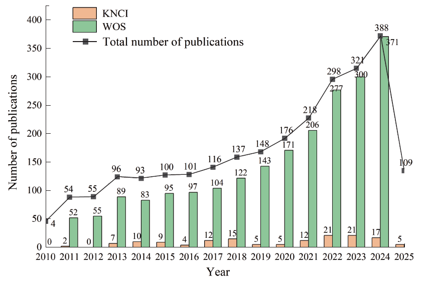
Mapping Global Research on Sustainable Textile Materials: A Comparative Bibliometric Study of China and International Literature
Cui-Yun Zhu , An-Ding Liu
View
A Lightweight Diffusion Framework for Cultural Pattern Generation Using LoRA
Jin-song Wang, Xiao-yu Xin, Shuai-shuai Qi, Ge-li Qin
View
Highlights
More
Special Issues
More
Most Read
Most Download
Useful Links
More